Anti-CXCR4 (EL) Antibody (1012)
$445.00
SKU: 1012
Categories: Antibody Products, Infectious Disease Antibodies, Products
Overview
Product Name Anti-CXCR4 (EL) Antibody (1012)
Description Anti-CXCR4 (EL) Rabbit Polyclonal Antibody
Target CXCR4 (EL)
Species Reactivity Human, Mouse
Applications ELISA,WB,IHC,IF
Host Rabbit
Clonality Polyclonal
Isotype IgG
Immunogen Peptide corresponding to aa 182- 196 in the second extracellular loop (EL) of human CXCR4.
Properties
Form Liquid
Concentration Lot Specific
Formulation PBS, pH 7.4.
Buffer Formulation Phosphate Buffered Saline
Buffer pH pH 7.4
Format Purified
Purification Purified by peptide immuno-affinity chromatography
Specificity Information
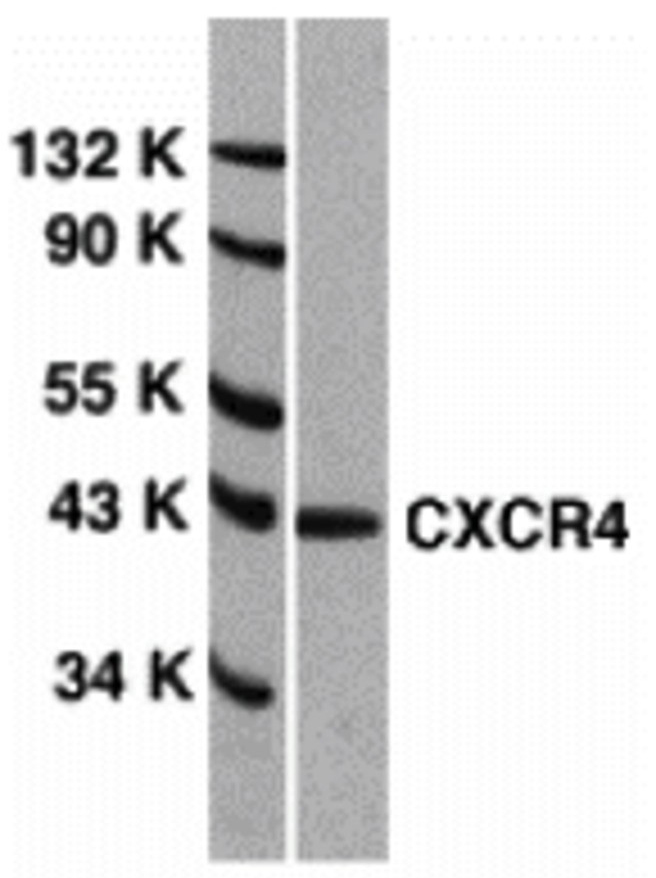
Specificity This antibody recognizes human and mouse CXCR4 (40 kD).
Target Name C-X-C chemokine receptor type 4
Target ID CXCR4 (EL)
Uniprot ID P61073
Alternative Names CXC-R4, CXCR-4, FB22, Fusin, HM89, LCR1, Leukocyte-derived seven transmembrane domain receptor, LESTR, Lipopolysaccharide-associated protein 3, LAP-3, LPS-associated protein 3, NPYRL, Stromal cell-derived factor 1 receptor, SDF-1 receptor, CD antigen CD184
Gene Name CXCR4
Gene ID 7852
Accession Number NP_003458
Sequence Location Cell membrane, Cell junction. Early endosome. Late endosome. Lysosome. Note=In unstimulated cells, diffuse pattern on plasma membrane. On agonist stimulation, colocalizes with ITCH at the plasma membrane where it becomes ubiquitinated. In the presence of antigen, distributes to the immunological synapse forming at the T-cell-APC contact area, where it localizes at the peripheral and distal supramolecular activation cluster (SMAC).
Biological Function Receptor for the C-X-C chemokine CXCL12/SDF-1 that transduces a signal by increasing intracellular calcium ion levels and enhancing MAPK1/MAPK3 activation (PubMed:10452968, PubMed:28978524, PubMed:18799424, PubMed:24912431). Involved in the AKT signaling cascade (PubMed:24912431). Plays a role in regulation of cell migration, e.g. during wound healing (PubMed:28978524). Acts as a receptor for extracellular ubiquitin; leading to enhanced intracellular calcium ions and reduced cellular cAMP levels (PubMed:20228059). Binds bacterial lipopolysaccharide (LPS) et mediates LPS-induced inflammatory response, including TNF secretion by monocytes (PubMed:11276205). Involved in hematopoiesis and in cardiac ventricular septum formation. Also plays an essential role in vascularization of the gastrointestinal tract, probably by regulating vascular branching and/or remodeling processes in endothelial cells. Involved in cerebellar development. In the CNS, could mediate hippocampal-neuron survival (By similarity). {UniProtKB:P70658, PubMed:10074102, PubMed:10452968, PubMed:10644702, PubMed:10825158, PubMed:11276205, PubMed:17197449, PubMed:18799424, PubMed:20048153, PubMed:20228059, PubMed:20505072, PubMed:24912431, PubMed:28978524, PubMed:8752280, PubMed:8752281}.; (Microbial infection) Acts as a coreceptor (CD4 being the primary receptor) for human immunodeficiency virus-1/HIV-1 X4 isolates and as a primary receptor for some HIV-2 isolates. Promotes Env-mediated fusion of the virus (PubMed:8849450, PubMed:8929542, PubMed:9427609, PubMed:10074122, PubMed:10756055). {PubMed:10074122, PubMed:10756055, PubMed:8849450, PubMed:8929542, PubMed:9427609}.
Research Areas Infectious Disease
Background Human immunodeficiency virus (HIV) and related viruses require coreceptors in addition to CD4 to infect target cells. Some G protein- coupled receptors in the chemokine receptor family, including CCR5, CXCR4, CCR3, CCR2b, and CCR8 in the chemokine receptor family, have been identified as HIV coreceptors. CXCR4 (also called Fusin, LESTR, HUMSTR) is a principal coreceptor for T-cell tropic strains of HIV-1. CXCR4 is also required for the infection by dual-tropic strains of HIV-1 and mediates CD4-independent infection by HIV-2. The alpha- chemokine SDF-1 is the ligand for EXCR4 and prevents infection by T-cell tropic HIV-1. CXCR4 associates with the surface CD4-gp120 complex before HIV enters target cells. The amino- terminal domain and the second extracellular loop of CXCR4 serve as HIV binding sites. CXCR4 messenger RNA levels correlate with HIV-1 permissiveness in diverse human cell types. Antibodies to CXCR4 may block HIV-1 and HIV-2 infection of human target cells.
Application Images


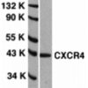
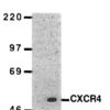
Description Western Blot Validation of CXCR4 in HeLa Cells
Loading: 15 ug of lysates per lane. Antibodies: 1012 (1 ug/mL), 1 h incubation at RT in 5% NFDM/TBST. Secondary: Goat anti-rabbit IgG HRP conjugate at 1:10000 dilution.
Loading: 15 ug of lysates per lane. Antibodies: 1012 (1 ug/mL), 1 h incubation at RT in 5% NFDM/TBST. Secondary: Goat anti-rabbit IgG HRP conjugate at 1:10000 dilution.

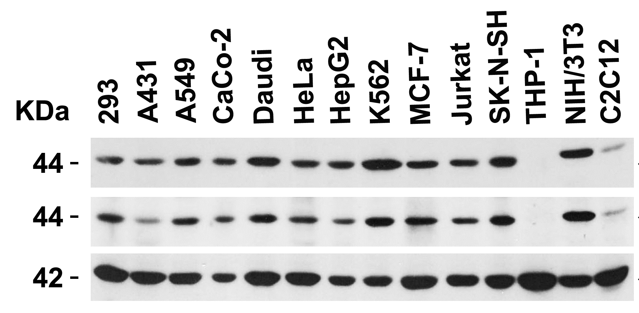
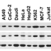
Description Independent Antibody Validation (IAV) via Protein Expression Profile
Loading: 15 ug of lysates per lane. Antibodies: 1009 (1 ug/mL), 1012 (1 ug/mL), and beta-actin (1 ug/mL), 1 h incubation at RT in 5% NFDM/TBST. Secondary: Goat anti-rabbit IgG HRP conjugate at 1:10000 dilution.
Loading: 15 ug of lysates per lane. Antibodies: 1009 (1 ug/mL), 1012 (1 ug/mL), and beta-actin (1 ug/mL), 1 h incubation at RT in 5% NFDM/TBST. Secondary: Goat anti-rabbit IgG HRP conjugate at 1:10000 dilution.

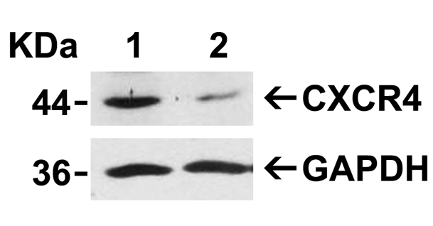
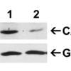
Description Validation with CXCR4 siRNA Knockdown in HeLa Cells
HeLa cells were transfected with control siRNAs (lane 1) or CXCR4 siRNAs (lane 2) Loading: 10 ug of HeLa whole cell lysates per lane. Antibodies: 1012 (2 ug/mL), 1 h incubation at RT in 5% NFDM/TBST. Secondary: Goat anti-rabbit IgG HRP conjugate at 1:10000 dilution.
HeLa cells were transfected with control siRNAs (lane 1) or CXCR4 siRNAs (lane 2) Loading: 10 ug of HeLa whole cell lysates per lane. Antibodies: 1012 (2 ug/mL), 1 h incubation at RT in 5% NFDM/TBST. Secondary: Goat anti-rabbit IgG HRP conjugate at 1:10000 dilution.

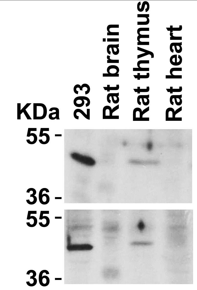
Description Animal Species Reactivity
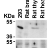
Loading: Lysates/proteins at 20 ug per lane. Antibodies: 1009 (2 ug/mL) or 1012 (2 ug/mL). 1 h incubation at RT in 5% NFDM/TBST.Secondary: Goat anti-rabbit IgG HRP conjugate at 1:10000 dilution.
Loading: Lysates/proteins at 20 ug per lane. Antibodies: 1009 (2 ug/mL) or 1012 (2 ug/mL). 1 h incubation at RT in 5% NFDM/TBST.Secondary: Goat anti-rabbit IgG HRP conjugate at 1:10000 dilution.

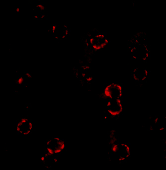
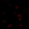
Description Immunofluorescence Validation of CXCR4
Immunofluorescent analysis of 4% paraformaldehyde-fixed HeLa cells labeling CXCR4 with 1012 at 4 ug/mL, followed by goat anti-rabbit IgG secondary antibody at 1/500 dilution (red). Image showing both membrane and cytoplasmic staining on HeLa cell line.
Immunofluorescent analysis of 4% paraformaldehyde-fixed HeLa cells labeling CXCR4 with 1012 at 4 ug/mL, followed by goat anti-rabbit IgG secondary antibody at 1/500 dilution (red). Image showing both membrane and cytoplasmic staining on HeLa cell line.

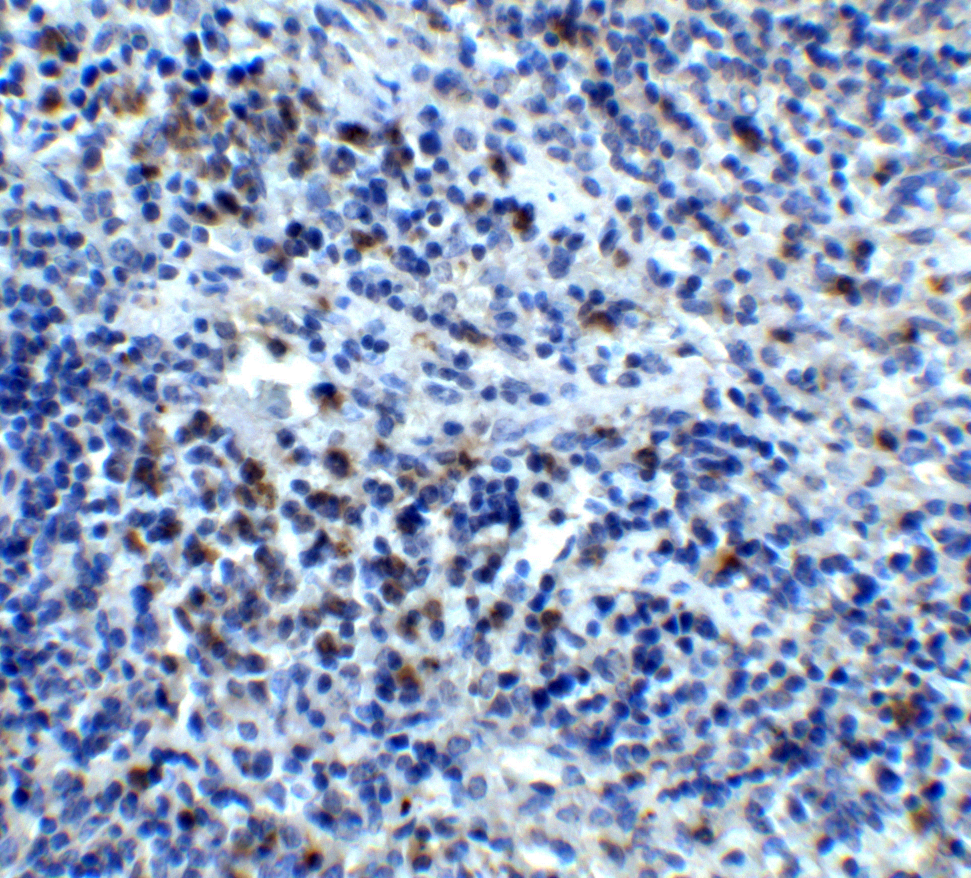
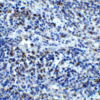
Description Immunohistochemistry Validation of CXCR4 in Human Spleen
Immunohistochemical analysis of paraffin-embedded human spleen tissue using anti-CXCR4 antibody (1012) at 5 ug/ml. Tissue was fixed with formaldehyde and blocked with 10% serum for 1 h at RT; antigen retrieval was by heat mediation with a citrate buffer (pH6). Samples were incubated with primary antibody overnight at 4 C. A goat anti-rabbit IgG H&L (HRP) at 1/250 was used as secondary. Counter stained with Hematoxylin.
Immunohistochemical analysis of paraffin-embedded human spleen tissue using anti-CXCR4 antibody (1012) at 5 ug/ml. Tissue was fixed with formaldehyde and blocked with 10% serum for 1 h at RT; antigen retrieval was by heat mediation with a citrate buffer (pH6). Samples were incubated with primary antibody overnight at 4 C. A goat anti-rabbit IgG H&L (HRP) at 1/250 was used as secondary. Counter stained with Hematoxylin.

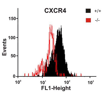
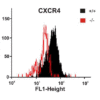
Description KO Validation of CXCR4 by Flow Cytometry (Ödemis, et al., 2010)
Astrocytes from wild-type or CXCR4 knockout mice were stained with primary antibodies against CXCR4 and FITC-labeled secondary antibodies, and subsequently subjected to flow cytometry. CXCR4−/− astrocytes (red) showed loss of CXCR4 cell-surface expression compared with wild-type cells (black).
Astrocytes from wild-type or CXCR4 knockout mice were stained with primary antibodies against CXCR4 and FITC-labeled secondary antibodies, and subsequently subjected to flow cytometry. CXCR4−/− astrocytes (red) showed loss of CXCR4 cell-surface expression compared with wild-type cells (black).

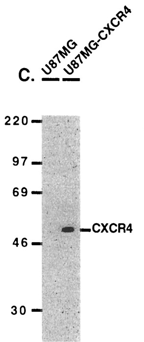
Description Overexpression Validation of CXCR4 (Kozak et al., 2002)
U87MG and U87MG-CXCR4 extracts were included as negative and positive controls, respectively, for CXCR4 detection with anti-CXCR4 antibodies.
U87MG and U87MG-CXCR4 extracts were included as negative and positive controls, respectively, for CXCR4 detection with anti-CXCR4 antibodies.

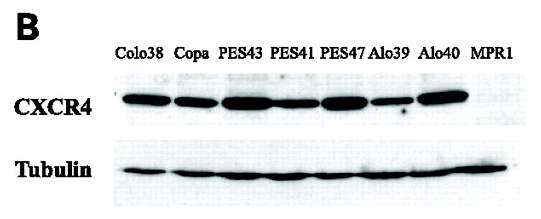
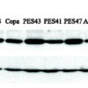
Description WB Validation of CXCR4 in Human Metastatic Melanoma (Scala et al., 2006)
CXCR4 protein was detected in the human metastatic melanoma cell lines and human melanoma cell line (colo38), but not in the human primary melanocytes (MPR1) with anti-CXCR4 antibodies.
CXCR4 protein was detected in the human metastatic melanoma cell lines and human melanoma cell line (colo38), but not in the human primary melanocytes (MPR1) with anti-CXCR4 antibodies.
Handling
Storage This antibody is stable for at least one (1) year at -20°C. Avoid multiple freeze-thaw cycles.
Dilution Instructions Dilute in PBS or medium which is identical to that used in the assay system.
Application Instructions Immunoblotting: use at 1:500-1:1,000 dilution.
Positive control: Whole cell lysate from HeLa cells.
Positive control: Whole cell lysate from HeLa cells.
References & Data Sheet
Data Sheet  Download PDF Data Sheet
 Download PDF Data Sheet

