Anti-EndoG Antibody (11004)
$445.00
| Host | Quantity | Applications | Species Reactivity | Data Sheet | |
|---|---|---|---|---|---|
| Rabbit | 100ug | ELISA,WB,IHC-P,IF | Human, Mouse, Rat |  |
SKU: 11004
Categories: Antibody Products, Apoptosis Antibodies, Products
Overview
Product Name Anti-EndoG Antibody (11004)
Description Anti-EndoG Rabbit Polyclonal Antibody
Target EndoG
Species Reactivity Human, Mouse, Rat
Applications ELISA,WB,IHC-P,IF
Host Rabbit
Clonality Polyclonal
Isotype IgG
Immunogen Peptide corresponding to aa 55- 70 of human EndoG (accession no. NP_004426).
Properties
Form Liquid
Concentration Lot Specific
Formulation PBS, pH 7.4.
Buffer Formulation Phosphate Buffered Saline
Buffer pH pH 7.4
Format Purified
Purification Purified by peptide immuno-affinity chromatography
Specificity Information
Specificity This antibody recognizes human, mouse, and rat EndoG (Endonuclease G) an enzyme that is activated by apoptotic stimuli and is able to induce nucleosomal fragmentation of DNA independently of caspase and DFF/CAD. EndoG is a mitochondria-specific nuclease that translocates to the nucleus and cleaves chromatin DNA during apoptosis.
Target Name Endonuclease G, mitochondrial
Target ID EndoG
Uniprot ID Q14249
Alternative Names Endo G, EC 3.1.30.-
Gene Name ENDOG
Gene ID 2021
Accession Number NP_004426
Sequence Location Mitochondrion
Biological Function Endonuclease that preferentially catalyzes the cleavage of double-stranded 5-hydroxymethylcytosine (5hmC)-modified DNA (PubMed:25355512). The 5hmC-modified nucleotide does not increase the binding affinity, but instead increases the efficiency of cutting and specifies the site of cleavage for the modified DNAs (By similarity). Shows significantly higher affinity for four-stranded Holliday junction over duplex and single-stranded DNAs (By similarity). Promotes conservative rUniProtKB:O08600, PubMed:25355512, PubMed:29719607, PubMed:33473107}.
Research Areas Apoptosis
Application Images


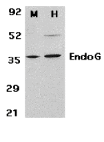
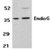
Description Western Blot Validation in Mouse 3T3 (M) and Human HepG2 (H) Cell Lysates
Loading: 15 ug of lysates per lane. Antibodies: EndoG 11004 (2 ug/mL), 1h incubation at RT in 5% NFDM/TBST.Secondary: Goat anti-rabbit IgG HRP conjugate at 1:10000 dilution.
Loading: 15 ug of lysates per lane. Antibodies: EndoG 11004 (2 ug/mL), 1h incubation at RT in 5% NFDM/TBST.Secondary: Goat anti-rabbit IgG HRP conjugate at 1:10000 dilution.

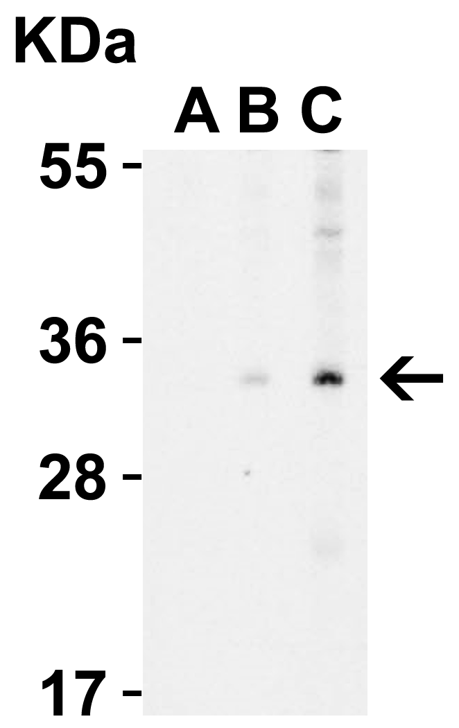
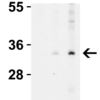
Description Western Blot Validation in Human A431 Cell Lysate with the presence (A) or absence (B and C) of blocking peptide
Loading: 15 ug of lysates per lane. Antibodies: EndoG 11004 (A: 0.5 ug/mL, B: 0.5 ug/mL, C: 1 ug/mL), 1h incubation at RT in 5% NFDM/TBST.Secondary: Goat anti-rabbit IgG HRP conjugate at 1:10000 dilution.
Loading: 15 ug of lysates per lane. Antibodies: EndoG 11004 (A: 0.5 ug/mL, B: 0.5 ug/mL, C: 1 ug/mL), 1h incubation at RT in 5% NFDM/TBST.Secondary: Goat anti-rabbit IgG HRP conjugate at 1:10000 dilution.

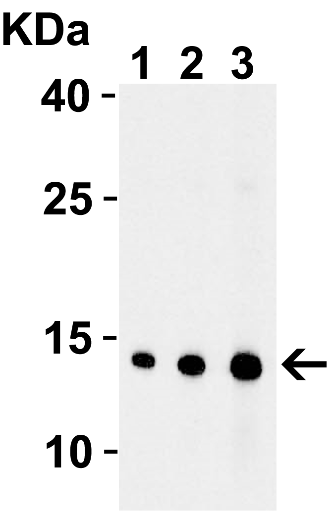
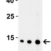
Description Western Blot Validation with Recombinant Protein
Loading: 30 ng of human EndoG recombinant protein per lane. Antibodies: EndoG 11004, 1h incubation at RT in 5% NFDM/TBST.Secondary: Goat anti-rabbit IgG HRP conjugate at 1:10000 dilution.Lane 1: 0.5 ug/mLLane 2: 1 ug/mLLane 3: 2 ug/mL
Loading: 30 ng of human EndoG recombinant protein per lane. Antibodies: EndoG 11004, 1h incubation at RT in 5% NFDM/TBST.Secondary: Goat anti-rabbit IgG HRP conjugate at 1:10000 dilution.Lane 1: 0.5 ug/mLLane 2: 1 ug/mLLane 3: 2 ug/mL

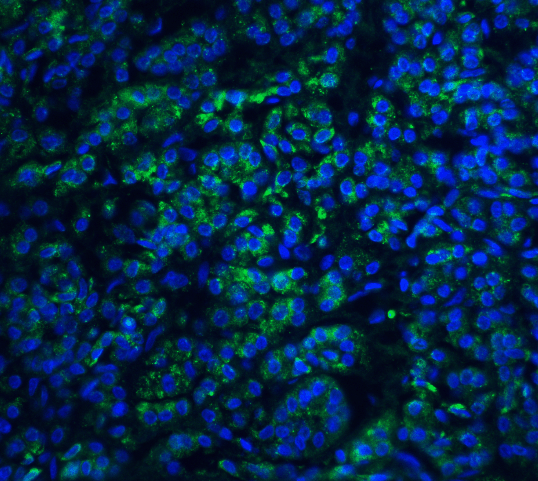
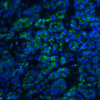
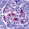
Description Immunofluorescence Validation of EndoG in Human Pancreas Tissue
Immunofluorescent analysis of 4% paraformaldehyde-fixed human pancreas tissue labeling EndoG with 11004 at 20 ug/mL, followed by goat anti-rabbit IgG secondary antibody at 1/500 dilution (green) and DAPI staining (blue).
Immunofluorescent analysis of 4% paraformaldehyde-fixed human pancreas tissue labeling EndoG with 11004 at 20 ug/mL, followed by goat anti-rabbit IgG secondary antibody at 1/500 dilution (green) and DAPI staining (blue).

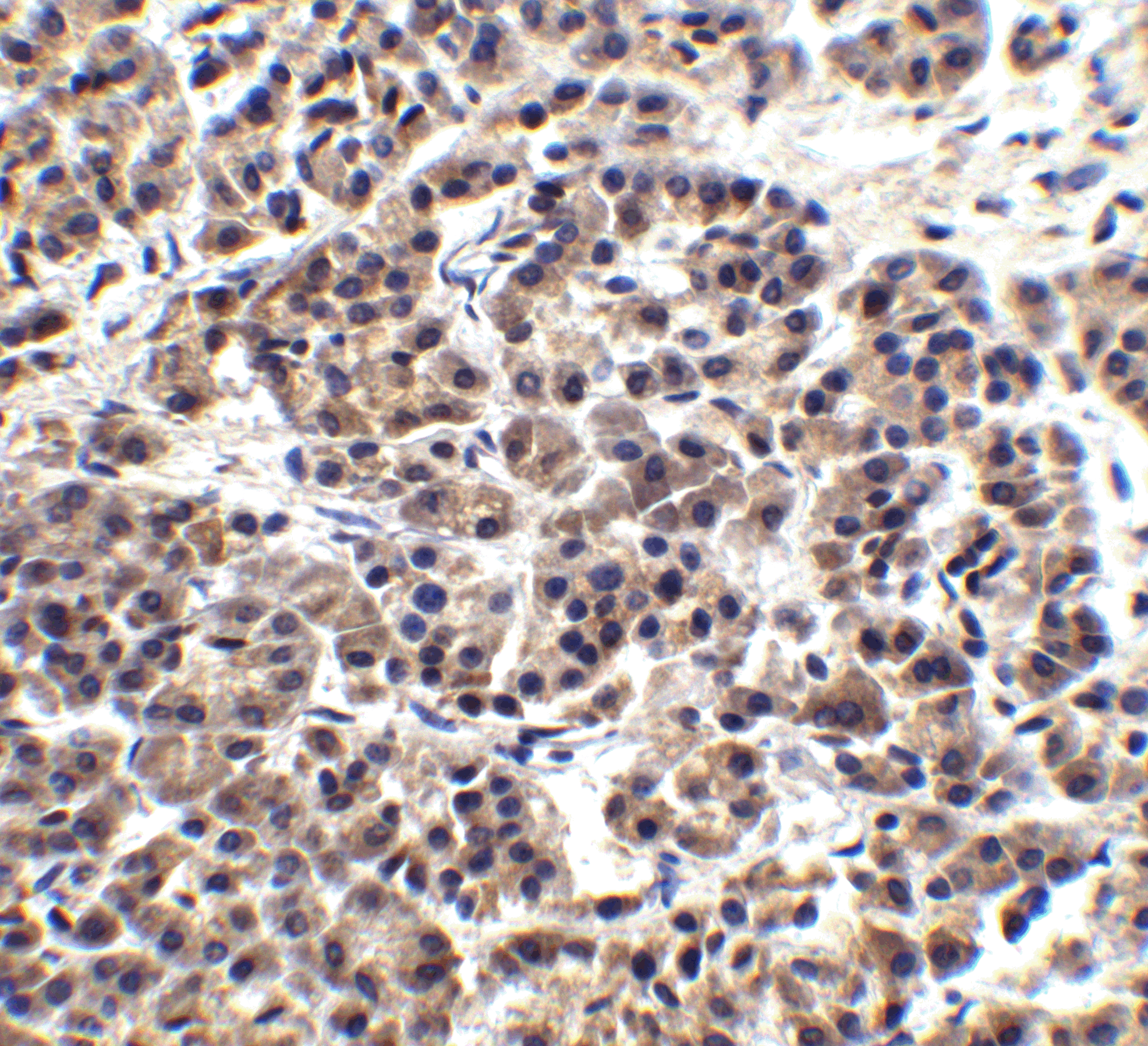
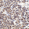
Description Immunohistochemistry Validation of EndoG in Human Pancreas Tissue
Immunohistochemical analysis of paraffin-embedded human pancreas tissue using anti-EndoG antibody (11004) at 15 ug/ml. Tissue was fixed with formaldehyde and blocked with 10% serum for 1 h at RT; antigen retrieval was by heat mediation with a citrate buffer (pH6). Samples were incubated with primary antibody overnight at 4C. A goat anti-rabbit IgG H&L (HRP) at 1/250 was used as secondary. Counter stained with Hematoxylin.
Immunohistochemical analysis of paraffin-embedded human pancreas tissue using anti-EndoG antibody (11004) at 15 ug/ml. Tissue was fixed with formaldehyde and blocked with 10% serum for 1 h at RT; antigen retrieval was by heat mediation with a citrate buffer (pH6). Samples were incubated with primary antibody overnight at 4C. A goat anti-rabbit IgG H&L (HRP) at 1/250 was used as secondary. Counter stained with Hematoxylin.

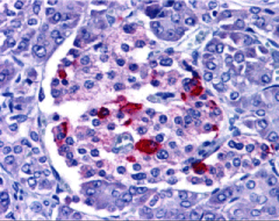
Description Immunohistochemistry Validation of EndoG in Human Pancreas Tissue
Immunohistochemical analysis of paraffin-embedded human pancreas tissue using anti-EndoG antibody (11004) at 2.5 ug/ml. Tissue was fixed with formaldehyde and blocked with 10% serum for 1 h at RT; antigen retrieval was by heat mediation with a citrate buffer (pH6). Samples were incubated with primary antibody overnight at 4C. A goat anti-rabbit IgG H&L (HRP) at 1/250 was used as secondary. Counter stained with Hematoxylin.
Immunohistochemical analysis of paraffin-embedded human pancreas tissue using anti-EndoG antibody (11004) at 2.5 ug/ml. Tissue was fixed with formaldehyde and blocked with 10% serum for 1 h at RT; antigen retrieval was by heat mediation with a citrate buffer (pH6). Samples were incubated with primary antibody overnight at 4C. A goat anti-rabbit IgG H&L (HRP) at 1/250 was used as secondary. Counter stained with Hematoxylin.

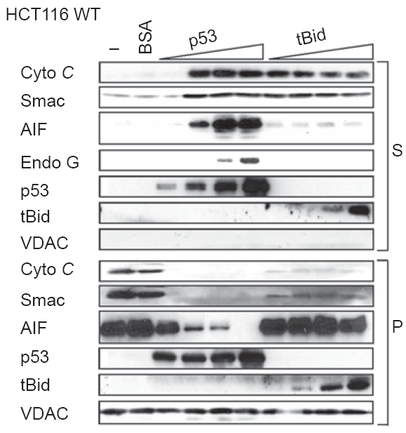
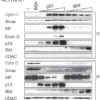
Description Localization Validation of EndoG by Purified p53 in Human Colorectal Cancer (HCT116) WT Cells (Wolff et al., 2008)
Immunoblot analysis of subcellular fraction enriched with supernatant (S) was used to determine EndoG protein levels with increasing amounts of p53 (10, 20, 40, 100nM) in HCT116 WT cells. The release of EndoG from mitochondria induced by p53 is detected by anti-EndoG antibodies.
Immunoblot analysis of subcellular fraction enriched with supernatant (S) was used to determine EndoG protein levels with increasing amounts of p53 (10, 20, 40, 100nM) in HCT116 WT cells. The release of EndoG from mitochondria induced by p53 is detected by anti-EndoG antibodies.

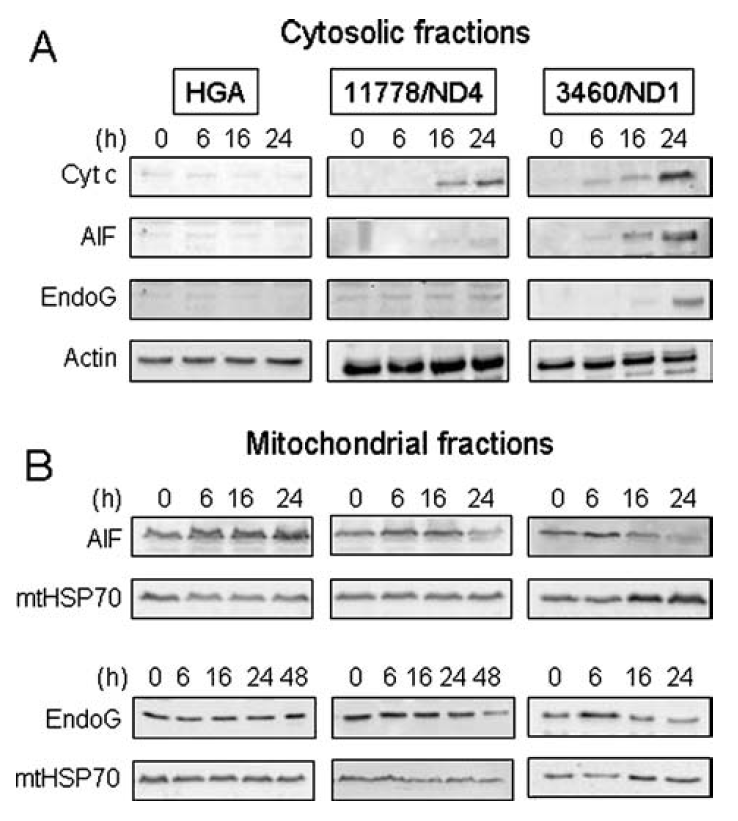
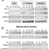
Description Localization Validation of EndoG in LHON cybrids (Zanna et al., 2005)
Immunoblots of subcellular fractions enriched for (A) cytosol and (B) mitochondria were used to determine EndoG protein levels with anti-EndoG antibodies in LHON cybrids. The release of EndoG from mitochondria into the cytosol of 3460/ND1 mutant is observed after 24hr. Control (HGA) was unaffected.
Immunoblots of subcellular fractions enriched for (A) cytosol and (B) mitochondria were used to determine EndoG protein levels with anti-EndoG antibodies in LHON cybrids. The release of EndoG from mitochondria into the cytosol of 3460/ND1 mutant is observed after 24hr. Control (HGA) was unaffected.

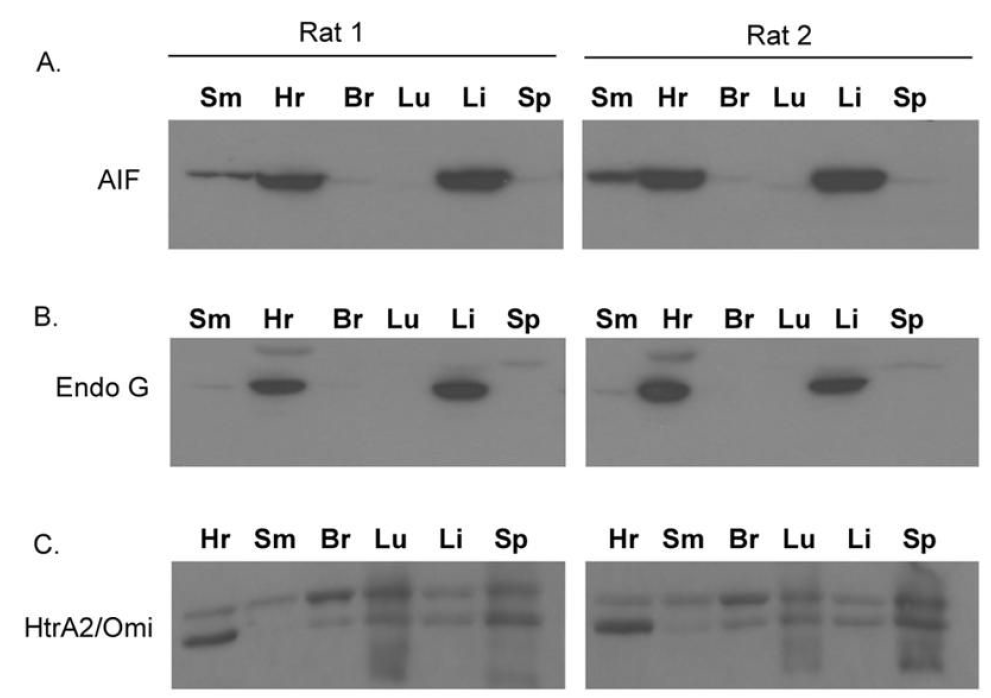
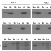
Description Tissue Specificity of EndoG in Rat Organs ( Siu et al., 2416)
WB analysis with anti-EndoG antibodies was performed for (B) EndoG in different organs of rats. EndoG was expressed very high in the heart and the liver of rats.
WB analysis with anti-EndoG antibodies was performed for (B) EndoG in different organs of rats. EndoG was expressed very high in the heart and the liver of rats.

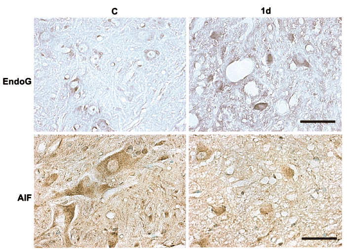
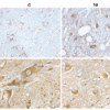
Description Immunohistochemistry Validation of EndoG translocation in Rat Spinal Cord Tissue (Yu et al., 2006)
Protein analysis for EndoG translocation by immunohistochemistry with anti-EndoG antibodies in Rat spinal cord tissue. The staining showed that EndoG was expressed in the nuclei of SCI rats (1d) as compared to the perikarya (cytoplasm) in WT.
Protein analysis for EndoG translocation by immunohistochemistry with anti-EndoG antibodies in Rat spinal cord tissue. The staining showed that EndoG was expressed in the nuclei of SCI rats (1d) as compared to the perikarya (cytoplasm) in WT.

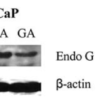
Description Translocation of EndoG by α-chaconine (CHA) and gallic acid (GA) in Human Prostate Cancer Cells (Reddivari et al., 2010)
Lymph-node carcinoma of the prostate (LNCaP) or prostate cancer-3 (PC-3) cells were treated with CHA (2.5 ug/ml) or GA (15 ug/ml) for 24hr. EndoG expression detected by anti-EndoG antibodies was increased significantly in nuclear fraction after CHA or GA treatments while this effect was not observed in the solvent control (C).
Lymph-node carcinoma of the prostate (LNCaP) or prostate cancer-3 (PC-3) cells were treated with CHA (2.5 ug/ml) or GA (15 ug/ml) for 24hr. EndoG expression detected by anti-EndoG antibodies was increased significantly in nuclear fraction after CHA or GA treatments while this effect was not observed in the solvent control (C).

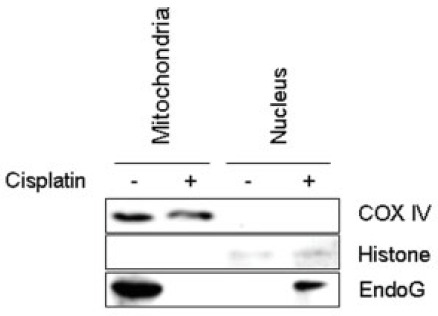
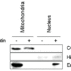
Description Translocation of EndoG by Cisplatin in Human HN4 cells (Kim et al., 2008)
Head and Neck Squamous Cell Carcinoma(HNSCC), HN4 cells, from the patients were treated with 40 μM cisplatin. An increased expression of EndoG was induced in the nuclear fraction by cisplatin treatment, which was not observed in the mitochondria.
Head and Neck Squamous Cell Carcinoma(HNSCC), HN4 cells, from the patients were treated with 40 μM cisplatin. An increased expression of EndoG was induced in the nuclear fraction by cisplatin treatment, which was not observed in the mitochondria.
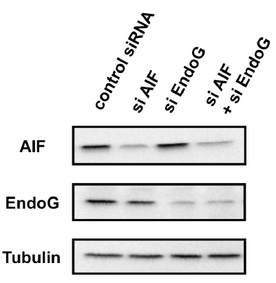
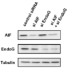
Description KD Validation of EndoG in Human small cell lung carcinoma Ms-1/Bcl-xL Cells (Sasazawa et al., 2009)
Western blot analysis with anti-EndoG antibodies was performed for EndoG in Ms-1/Bcl-xL cells transfected with EndoG siRNA or control siRNA. EndoG expression was downregulated after EndoG siRNA knockdown.
Western blot analysis with anti-EndoG antibodies was performed for EndoG in Ms-1/Bcl-xL cells transfected with EndoG siRNA or control siRNA. EndoG expression was downregulated after EndoG siRNA knockdown.
Handling
Storage This antibody is stable for at least one (1) year at -20°C. Avoid multiple freeze-thaw cycles.
Dilution Instructions Dilute in PBS or medium which is identical to that used in the assay system.
Application Instructions Immunoblotting: use at 1-2ug/mL. A band of 35kDa is detected.
Immunohistochemistry: use at 10-15ug/mL.
Positive control: HepG2 cell lysate.
These are recommended concentrations.
Enduser should determine optimal concentrations for their applications
Immunohistochemistry: use at 10-15ug/mL.
Positive control: HepG2 cell lysate.
These are recommended concentrations.
Enduser should determine optimal concentrations for their applications
References & Data Sheet
Data Sheet  Download PDF Data Sheet
 Download PDF Data Sheet

