Anti-BNIP3 Antibody (2290)
$487.00
SKU: 2290
Categories: Antibody Products, Apoptosis Antibodies, Products
Overview
Product Name Anti-BNIP3 Antibody (2290)
Description Anti-BNIP3 Rabbit Polyclonal Antibody
Target BNIP3
Species Reactivity Human, Mouse
Applications WB,ICC/IF
Host Rabbit
Clonality Polyclonal
Immunogen Synthetic peptide corresponding to amino acids at the C-terminus of human BNIP3.
Properties
Form Liquid
Concentration 1.0 mg/mL
Formulation PBS, pH 7.4, 50% glycerol, 0.09% sodium azide.
Buffer Formulation Phosphate Buffered Saline
Buffer pH pH 7.4
Buffer Anti-Microbial 0.09% Sodium Azide
Buffer Cryopreservative 50% Glycerol
Format Purified
Purification Purified by peptide immuno-affinity chromatography
Specificity Information
Specificity This antibody recognizes human and mouse BNIP3.
Target Name BCL2/adenovirus E1B 19 kDa protein-interacting protein 3
Target ID BNIP3
Uniprot ID Q12983
Gene Name BNIP3
Gene ID 664
Accession Number NP_004043.3
Sequence Location Mitochondrion. Mitochondrion outer membrane; Single-pass membrane protein. Note=Coexpression with the EIB 19-kDa protein results in a shift in NIP3 localization pattern to the nuclear envelope. Colocalizes with ACAA2 in the mitochondria. Colocalizes with SPATA18 at the mitochondrion outer membrane.
Biological Function Apoptosis-inducing protein that can overcome BCL2 suppression. May play a role in repartitioning calcium between the two major intracellular calcium stores in association with BCL2. Involved in mitochondrial quality control via its interaction with SPATA18/MIEAP: in response to mitochondrial damage, participates in mitochondrial protein catabolic process (also named MALM) leading to the degradation of damaged proteins inside mitochondria. The physical interaction of SPATA18/MIEAP, BNIP3 and BNIP3L/NIX at the mitochondrial outer membrane regulates the opening of a pore in the mitochondrial double membrane in order to mediate the translocation of lysosomal proteins from the cytoplasm to the mitochondrial matrix. Plays an important role in the calprotectin (S100A8/A9)-induced cell death pathway. {PubMed:19935772, PubMed:22292033}.
Research Areas Apoptosis
Background BNIP3 is a member of the apoptotic Bcl-2 protein family that is involved in both necrosis and apoptosis. Humans and other animals, as well as lower eukaryotes, encode several BNIP3 paralogues including human BNIP3L which induces apoptosis by interacting with viral and cellular anti-apoptosis proteins. BNIP3 interacts with the E1B 19kDa protein, which is responsible for the protection of virally induced cell death, as well as with E1B-like sequences of BCL2 which is also an apoptotic protector. The BNIP3 gene contains a BH3 domain and a transmembrane domain that have been associated with pro-apoptotic function. The dimeric mitochondrial protein encoded by the BNIP3 gene is known to induce apoptosis, even in the presence of BCL2.
Application Images


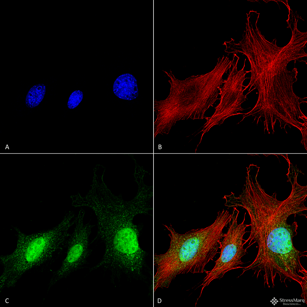
Description Immunocytochemistry/Immunofluorescence analysis using Rabbit Anti-BNIP3 Polyclonal Antibody (2290). Tissue: Fibroblast cell line (NIH 3T3). Species: Mouse. Fixation: 4% Formaldehyde for 15 min at RT. Primary Antibody: Rabbit Anti-BNIP3 Polyclonal Antibody (2290) at 1:100 for 60 min at RT. Secondary Antibody: Goat Anti-Rabbit ATTO 488 at 1:100 for 60 min at RT. Counterstain: Phalloidin Texas Red F-Actin stain; DAPI (blue) nuclear stain at 1:1000, 1:5000 for 60min RT, 5min RT. Localization: Cytoplasm . Magnification: 60X. (A) DAPI (blue) nuclear stain (B) Phalloidin Texas Red F-Actin stain (C) BNIP3 Antibody (D) Composite.

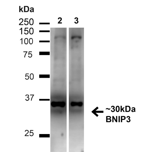
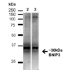
Description Western blot analysis of Human HeLa and HEK293T cell lysates showing detection of ~30kDa BNIP3 protein using Rabbit Anti-BNIP3 Polyclonal Antibody (2290). Lane 1: MW Ladder. Lane 2: Human HeLa (20 µg). Lane 3: Human 293T (20 µg). Load: 20 µg. Block: 5% milk + TBST for 1 hour at RT. Primary Antibody: Rabbit Anti-BNIP3 Polyclonal Antibody (2290) at 1:1000 for 1 hour at RT. Secondary Antibody: Goat Anti-Rabbit: HRP at 1:2000 for 1 hour at RT. Color Development: TMB solution for 12 min at RT. Predicted/Observed Size: ~30kDa .

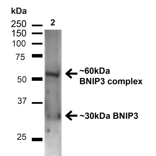
Description Western blot analysis of Mouse Kidney showing detection of ~30kDa BNIP3 protein using Rabbit Anti-BNIP3 Polyclonal Antibody (2290). Lane 1: MW Ladder. Lane 2: Mouse Kidney (20 µg). Load: 20 µg. Block: 5% milk + TBST for 1 hour at RT. Primary Antibody: Rabbit Anti-BNIP3 Polyclonal Antibody (2290) at 1:1000 for 1 hour at RT. Secondary Antibody: Goat Anti-Rabbit: HRP at 1:2000 for 1 hour at RT. Color Development: TMB solution for 12 min at RT. Predicted/Observed Size: ~30kDa . Other Band(s): ~60kDa (BNIP3 complex).
Handling
Storage This antibody is stable for at least one (1) year at -20°C.
Dilution Instructions Dilute in PBS or medium which is identical to that used in the assay system.
Application Instructions Immunoblotting: use at 1ug/mL. A band of ~30kDa is detected; an additional ~60kDa band, complexed BNIP3, may also be detected.
Immunofluorescence: use at 10ug/mL.
These are recommended concentrations.
Endusers should determine optimal concentrations for their applications.
Immunofluorescence: use at 10ug/mL.
These are recommended concentrations.
Endusers should determine optimal concentrations for their applications.
References & Data Sheet
Data Sheet  Download PDF Data Sheet
 Download PDF Data Sheet

