Anti-β-Actin Antibody (56574)
$466.00
SKU: 56574
Categories: Antibody Products, Neuroscience and Signal Transduction Antibodies, Products
Overview
Product Name Anti-β-Actin Antibody (56574)
Description Anti-β-Actin Rabbit Polyclonal Antibody
Target β-Actin
Species Reactivity Human
Applications WB,ICC/IF
Host Rabbit
Clonality Polyclonal
Immunogen Synthetic peptide corresponding to mid-protein amino acids of human beta-actin.
Properties
Form Liquid
Concentration 1.0 mg/mL
Formulation PBS, pH 7.4, 50% glycerol, 0.09% sodium azide.
Buffer Formulation Phosphate Buffered Saline
Buffer pH pH 7.4
Buffer Anti-Microbial 0.09% Sodium Azide
Buffer Cryopreservative 50% Glycerol
Format Purified
Purification Purified by Protein A affinity chromatography
Specificity Information
Specificity This antibody recognizes human beta-actin.
Target Name Actin, cytoplasmic 1
Target ID β-Actin
Uniprot ID P60709
Alternative Names β-actin [Cleaved into: Actin, cytoplasmic 1, N-terminally processed]
Gene Name ACTB
Sequence Location Cytoplasm, cytoskeleton, Nucleus
Biological Function Actin is a highly conserved protein that polymerizes to produce filaments that form cross-linked networks in the cytoplasm of cells (PubMed:29581253). Actin exists in both monomeric (G-actin) and polymeric (F-actin) forms, both forms playing key functions, such as cell motility and contraction (PubMed:29581253). In addition to their role in the cytoplasmic cytoskeleton, G- and F-actin also localize in the nucleus, and regulate gene transcription and motility and repair of damaged DNA (PubMed:29925947). {PubMed:29581253, PubMed:29925947}.
Research Areas Neuroscience
Background Actins are essential components of the cytoskeleton with critical roles in a wide range of cellular processes including cell migration, cell division, and the regulation of gene expression. Beta-actin, product of the Actb gene, is one of six different actin isoforms and is one of the two nonmuscle cytoplasmic actins (gamma-actin is the other); both are ubiquitously expressed and highly conserved from birds to mammals. Mutations in the Actb gene have been associated with diffuse large B-cell lymphoma. Beta-actin (molecular weight ~42kDa) is often used as a loading control in Western blots.
Application Images


Description Immunocytochemistry/Immunofluorescence analysis using Rabbit Anti-Beta Actin Polyclonal Antibody (56574). Tissue: Cervical cancer cell line (HeLa). Species: Human. Fixation: 4% Formaldehyde for 15 min at RT. Primary Antibody: Rabbit Anti-Beta Actin Polyclonal Antibody (56574) at 1:100 for 60 min at RT. Secondary Antibody: Goat Anti-Rabbit ATTO 488 at 1:100 for 60 min at RT. Counterstain: DAPI (blue) nuclear stain at 1:5000 for 5 min RT. Localization: Cytoplasm, Cytoskeleton. Magnification: 40X. (A) DAPI (blue) nuclear stain (B) Phalloidin Texas Red F-Actin stain (C) Beta Actin Antibody (D) Composite.

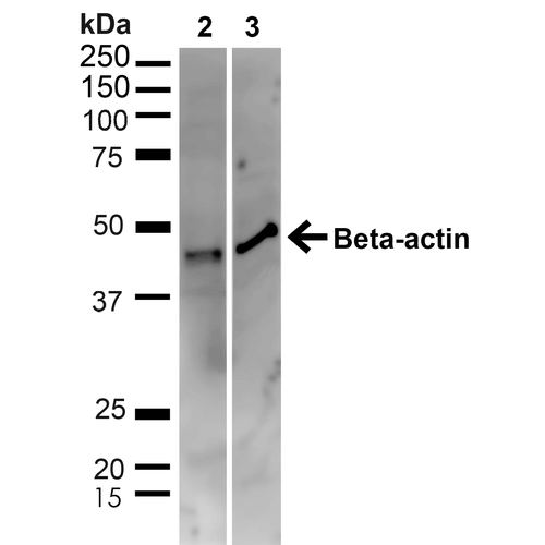
Description Western blot analysis of Human HeLa and HEK293T cell lysates showing detection of ~41.7kDa beta Actin protein using Rabbit Anti-beta Actin Polyclonal Antibody (56574). Lane 1: MW Ladder. Lane 2: Human HeLa (20 µg). Lane 3: Human 293T (20 µg). Load: 20 µg. Block: 5% milk + TBST for 1 hour at RT. Primary Antibody: Rabbit Anti-beta Actin Polyclonal Antibody (56574) at 1:1000 for 1 hour at RT. Secondary Antibody: Goat Anti-Rabbit: HRP at 1:2000 for 1 hour at RT. Color Development: TMB solution for 12 min at RT. Predicted/Observed Size: ~41.7kDa.
Handling
Storage This antibody is stable for at least one (1) year at -20°C.
Dilution Instructions Dilute in PBS or medium that is identical to that used in the assay system.
Application Instructions Immunoblotting: use at 1ug/mL.
Immunofluorescence: use at 10ug/mL.
These are recommended concentrations;
Endusers should determine optimal concentrations for their applications.
Immunofluorescence: use at 10ug/mL.
These are recommended concentrations;
Endusers should determine optimal concentrations for their applications.
References & Data Sheet
Data Sheet  Download PDF Data Sheet
 Download PDF Data Sheet


